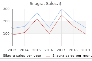
"Silagra 100mg without prescription, erectile dysfunction injections cost".
By: P. Tukash, M.B. B.A.O., M.B.B.Ch., Ph.D.
Deputy Director, Donald and Barbara School of Medicine at Hofstra/Northwell
It may help in the evaluation of hemodynamic effects of sternal closure gonorrhea causes erectile dysfunction buy silagra 50mg low cost, termination of ventricular assist devices or extracorporeal membrane oxygenation impotence over 60 purchase 100mg silagra visa. Post-bypass echo-Doppler evaluation is able to immediately assess the quality of the surgical repair and to assess cardiac function by examining ventricular wall motion and systolic thickening penile injections for erectile dysfunction side effects discount silagra 100 mg with mastercard. This technique can show residual structural defects after bypass erectile dysfunction doctor visit buy silagra 50 mg with visa, which can be immediately repaired in the same operative setting and prevent the patient from leaving the operating room with significant residual structural defects that later require reoperation. Importantly, post-bypass ventricular dysfunction and residual structural defects are identified by echo-Doppler assessment; left uncorrected, these are associated with an increased incidence of reoperation and greater morbidity and mortality. This monitoring tool helps assess surgical outcome and identify operative risk factors. Surgeons can demonstrate an operative learning curve with a reduced incidence of residual defects with experience. However, even when experienced surgeons perform the procedures, the use of an intraoperative echocardiogram can detect a 3 to 4 percent incidence of clinically significant residual disease that requires further surgical repair. Patients leaving the operating room with residual disease have a considerable increase in hospital cost, length of stay, and need for further operative or interventional procedures. Further, the use of an intraoperative echocardiogram can detect a 3 to 4 percent incidence of clinically significant residual disease that requires further surgical repair. Damage to the oropharynx, esophagus, brachial plexus, airway obstruction and various degree of dislodgement of the endotracheal tube have been reported. The probe related injuries include thermal injury, mechanical problems resulting in lacerations or perforation of the pharynx, hypopharynx, esophagus or stomach. Children with Down syndrome have intrinsic narrowing of the hypopharynx in addition to having an increased incidence of cervical spine narrowing that may result in difficult or failed probe placement. The first goal is to improve understanding of the cerebral function and dysfunction during cardiac surgery, so that effective brain protection strategies can be developed. For example, during deep hypothermia and before total circulatory arrest, the electroencephalogram can identify residual cerebral electrical activity. Because any residual electrical activity during arrest is associated with cerebral metabolism above basal activity, an isoelectric state may minimize ischemic injury to the brain during circulatory arrest. The use of drug-induced electrical silence does not have the same protective effect as hypothermia and may contribute to hemodynamic compromise in patients with postoperative myocardial dysfunction. In addition, the absence of electrical activity, particularly in newborns, does not necessarily correlate with optimal brain cooling. And may not be as useful in newborns as has been suggested in adults to ensure optimal cerebral protection from hypothermia. In newborns and infants, its reliability has been questioned, as processed electroencephalographic monitoring and its associated numerical correlation with anesthesia depth are based on adult electroencephalographic wave forms. Possible seizure activity in the postoperative period should be suspected when physiologic parameters such as tachycardia or hypertension are seen. A low threshold for electroencephalographic evaluation or the use of antiepileptic agents as part of postoperative sedation (midazolam) should be strongly considered in the neonatal population. Quantification of this important mechanism of cerebral injury during cardiac surgery would be instructive, because it has been suggested to be a contributor to neurologic injury. Marked differences between perfusion to the right or the left side have suggested problems with adequate surgical arterial or venous cannulation placement. In addition, low cerebral oxygen delivery can be inferred by reductions in the measured oxyhemoglobin saturation index levels. In addition to operative monitoring, there has been an increased interest in postoperative cerebral monitoring that may help determine adequate cerebral oxygenation trends. Some congenital cardiac centers have begun to use a noninvasive cerebral oxygen saturation monitor as an adjunct for trends in effective cardiac output and oxygen delivery. An additional advantage of this technique is the capability of assessing rapid alterations in blood flow velocity due to temperature or perfusion changes, as commonly occur during cardiac surgery. Selection of an induction technique is dependent on the degree of cardiac dysfunction, the cardiac defect, and the degree of sedation provided by the premedication. Other anesthetic considerations include a potential en hanced sensitivity to anesthetic drugs and correspondingly diminished anesthetic requirements, cardiomyopathy, cardiac conduction abnormalities and skeletal (specifically bulbar, oropharyngeal, and respiratory) muscle weakness. Reducing the doses, delayed onset, and greater time to recover from intravenous or inhaled agent, sedative, narcotic, and/or muscle relaxant should be anticipated. It is likely that many children will be more sensitive to and take more time to recover after non-depolarizing muscle relaxants, although here again, specific data are lacking. Few, if any, of these children (except the minority who have demonstrable multicore or minicore pathology) should be considered to be susceptible to malignant hyperthermia. The molecular pathophysiology of these disorders is quite different from that believed to underlie susceptibility to malignant hyperthermia, and there is little if any justification for avoiding known triggering agents on that basis. What is less clear is whether depolarizing muscle relaxants (and potentially inhaled agents such as halothane) have the ability in some children to induce sufficient and rapid skeletal muscle injury to provoke acute rhabdomyolysis and severe hyperkalemia (resulting in cardiac arrest, myoglobinuria, potential renal damage, etc. Given these considerations, controlled ventilation with endotracheal intubation is probably prudent for most circumstances given the potential for aspiration, airway obstruction, and respiratory muscle weakness. Full return to baseline level of consciousness, effort and strength are needed before extubation. The potentially increased and additive or synergistic effects of residual amounts of inhaled and intravenous agents must be appreciated, as should the potential for very prolonged (and seemingly idiosyncratic and/or related to the aforementioned changes in pharmacodynamics and pharmacokinetics) neuronal depression and delayed awakening. In children with good cardiac reserve, induction techniques can be quite varied as long as induction is careful and well 1032 monitored. The execution of induction is more important than the specific anesthetic technique in patients with reasonable cardiac reserve. A wide spectrum of anesthetic induction techniques with a variety of agents has been used safely and successfully; such as sevoflurane, sevoflurane and nitrous oxide, halothane, halothane and nitrous oxide, intravenous or intramuscular ketamine, or intravenous fentanyl, midazolam, propofol, dexmedetomidine or thiopental. In patients with more limited cardiac reserve, the choice of induction agent becomes more important.
Avoidance or reduction of myocardial edema occurs by limiting the pressure of cardioplegia infusions and by providing moderately hyperosmolar cardioplegia solutions that contain blood erectile dysfunction kolkata buy cheap silagra 100mg online. Close management of myocardial calcium balance to avoid extremes of intracellular hypercalcemia or hypocalcemia zantac causes erectile dysfunction generic silagra 100mg overnight delivery, especially during reperfusion erectile dysfunction doctor new orleans generic 100 mg silagra otc, is very important erectile dysfunction pills supplements purchase 50mg silagra mastercard. The addition of magnesium may solve this dilemma by preventing damage from higher cardioplegic calcium concentrations by its action as a calcium antagonist. This prevents mitochondrial calcium overload as a consequence of reperfusion injury. Magnesium also prevents the influx of sodium into the postischemic myocardium, which is exchanged for calcium during reperfusion. Every cardiac program has their own philosophy regarding cardioplegia and myocardial protection. In neonates and infants, albumin is added to the cardioplegic solution to maintain an appropriate colloid osmotic pressure. In children undergoing circulatory arrest, long crossclamp times, and large pump suction return cases, 20 mg/kg methylprednisolone is used up to a maximum of 500 mg, to reduce the production of inflammatory mediators that result in myocardial dysfunction. Mild degrees of hypothermia and certainly the avoidance of hyperthermia are essential in the perioperative period. Studies have shown that the temperature of the foot is more sensitive than the temperature of the hand and for anatomic or physiologic reasons, temperature gradients in the toes develop more readily than those in the fingers. Several end points have been proposed, such as nasopharyngeal temperatures greater than 35. If air is present, further deairing should occur before attempting to come off bypass. In the initial stages, after separating from bypass, additional volume can be administered by the perfusionist via the aortic cannula, usually under the direction of the surgeon or anesthesiologist. This involves taking arterial blood from the aortic cannula and passing this blood through the ultrafilter. Before this is done, both the perfusionist and the surgical team should be informed that protamine is about to be administered. The surgeons should remove any pump suckers from the field and the perfusionist should stop all pump suction. This is to ensure that no protamine enters the bypass circuit in case it is necessary to go back on bypass for any reason. Any blood products required are usually given after the administration of protamine, and these are usually given, while the surgeons are achieving hemostasis. As soon as the Rewarming Rewarming may have begun before release of the crossclamp, but more usually the child is rewarmed only after release of the clamp. After completion of the intracardiac repair and deairing of the heart, the aortic crossclamp is removed, allowing reperfusion of the myocardium. Optimally, normal sinus rhythm and myocardial contractility are restored during this time, while the child is slowly rewarmed. During rewarming, surgery is completed, inotropic and vasoactive agents are started, and ventilation resumes. Left atrial and/or pulmonary artery monitoring lines, if indicated, are placed at this time. Various degrees of heart block are common after heart surgery and it is most frequently associated with the administration of cardioplegia. As the heart is reperfused for a longer time, and the effects of cardioplegia are reduced, normal sinus rhythm is usually restored. However, heart block may result from damage to the conducting system during surgery and may require temporary atrial and ventricular pacing. Limiting protamine to this dose prevents overdosing of protamine with its associated effects on platelet function (reduction of the interaction of glycoprotein Ib receptor interaction with von Willebrand factor). However, particularly in infants, the administration of protamine and the persistent treatment of a suspected incomplete heparin reversal should not distract and delay the treatment of other commonly associated postbypass coagulopathies such as thrombocytopenia, platelet dysfunction, and other coagulation factor deficiencies. Protamine reactions are much less frequent in children younger than 16 years of age and are reported as 1. Independent risk factors are a female gender, a larger protamine dose, and smaller heparin doses. Type I reactions or effects during administration are rare and adding calcium does not change the hemodynamic consequences of injection. Administering the protamine over no less than 5 minutes reduces the severity and precipitous nature of any protamine reaction. Unstable neonates and small infants may have their sternums temporarily left open, with surgical closure planned 24 to 72 hours later when cardiac function has improved and myocardial edema diminished. In general, physiologic responses to bypass are more extreme with decreasing age and size of the child. The neonate experiences a greater degree of hemodilution on bypass and colder temperatures on bypass and frequently requires longer aortic cross-clamp times, all of which can result in a greater inflammatory response. The hemofilter has thousands of fibers with pores, which allow water, electrolytes and small molecules to be filtered out of the blood. This improves systolic and diastolic function of the myocardium and reduces endothelial dysfunction in the systemic and pulmonary vasculature. Clinically, however, any ultrafiltration method seems to benefit children, especially those undergoing complex repairs, neonates, and children with preexisting pulmonary hypertension. The disadvantages are that the child remains heparinized, body temperature may decrease during the process (unless the circuit is modified to include the heat exchanger). It requires extra time, an aortic cannula is needed that can obstruct the aorta in small infants, and acute intravascular volume shifts may occur at a time when the child is prone to hemodynamic instability.
Generic silagra 100mg free shipping. Sun News Dr.X - Premature EjaculationErectile dysfunction treatments Chennai ARC Research Centre.
Occasional patients have more than one granular cell tumor of the vulva erectile dysfunction injection therapy cost order silagra with amex, typically in association with similar tumors in extravulvar sites erectile dysfunction before 30 buy cheap silagra 50 mg on line. B: In this example erectile dysfunction exercise video effective silagra 100mg, small fascicles of spindle-shaped smooth muscle cells are separated by myxohyaline stroma erectile dysfunction pills dischem discount silagra 100 mg visa. A: the tumor has expanded and largely replaced the dermal compartment, and focally infiltrates subcutaneous adipose tissue (circlet:ll. Malignant granular cell tumors do exist, but their histologic features overlap with benign twnors and as a practical matter are generally not diagnosed as malignant until aggressive clinical behavior has become apparent. Tumors with large size, high mitotic rates, foci of necrosis, and/or spindle cell morphology should be flagged as tumors with an increased likelihood of possible malignant behavior. A: the architecture of 1he hyperplastic squamous epithelium, which includes formation of jagged nests. In order to arrive at the correct diagnosis, 1he underlying granular cell tumor must be recognized. B: Nests of pseudoepitheliomatous squamous epithelium are seen in intimate association with a granular cell tumor. Electron microscopic studies have shown that the numerous intracellular granules seen at the light microscopic level correspond to lysosomes. Nodular Fasciitis11 Although quite rare in the vulva, nodular fasciitis is presented in some detail due to the importance of its proper recogni~ tion in general surgical pathology. When it involves the vulva, this reactive proliferation of myofibroblasts usually occurs in the labial region of women of reproductive age, and has an average size of about 2. Histologically, the nodular proliferation often atends into neighboring adipose tissue for short distances. The lesional stromal cells consist of plump, mitotically active myo6broblasts with finely stippled chromatin and small nucleoli that are most often arranged in patterns that resemble myofibroblasts grown in tissue cultun: or found in granulation tissue. These myofibroblasts typically form short, curved bundles within a loose, feathery background that contains extravasated erytb~ rocytes and scattered lymphocytes. Mon: cellular regions with a fascicular, storiform, or sheedike architecture may also be present. Nodular fasciitis may also contain deposits of hya~ linized collagen and small aggregates of multinucleated giant cells associated with foci of mic:rohcmorrhage. At scanning magnification, 1hese lesions are seen to have a predominantly nodular configuration, but often extend into the adjacent adipose tissue for short distances. B: In this case, scattered osteoclast-like multinucleated giant cells are present in an area of microhemorrhage. With specific reference to vulvar soft tissue tumors and tumor-like lesions that are in the differential diagnosis, the postoperative spindle cell nodule and aggressive angiomy. As discussed in Chapter 2, postoperative spindle cell nodules are associated with a previous recent operative procedure at the site of the lesion. Although they share many histologic features with nodular fasciitis, they have a distinctive clinical history, tend to be more uniformly cellular and less varied in. Misinterpreting postoperative spindle cell nodule as nodular fasciitis, or vice versa. In comparison with nodular fasciitis, aggressive angiomyxoma is usually a larger mass, has a much more distinctive and prominent vascular pattern with widely patent vessel lumens, is less cellular, and its stromal cells are mitotica11y inactive with inconspicuous nucleoli. Epithelioid sarcomas are of unknown histogenesis, and have an immunopbenotype that characteristically includes coexpression of cytokeracin, epithelial membrane antigen, and vimentin. At scanning magnification, 1he multinodular growlh pattern and randomly distributed foci of tumor necrosis are evident in this proximal-type tumor. The reader is referred to the seminal anicle on proximal-type epithelioid sarcoma for a more in-depth discussion of the differential diagnosis of this unusual tumor. The cut surface of this subcutaneous nodule has a variegated appearance and contains foci of hemorrhage and necrosis. The lower 1hird of the epithelium exhibits nuclear atypia and disordered maturation, while koilocytes predominate in 1he upper portion.
The lumen is slightly dilated erectile dysfunction doctors in houston tx cheap silagra 50mg overnight delivery, the plical architecture varies from blunted and simplified to flattened prostate cancer erectile dysfunction statistics buy cheap silagra 50mg online, and the lamina propria of the few residual polypoid fronds is chronically inflamed erectile dysfunction drug has least side effects generic 100 mg silagra with mastercard. Ifa fallopian tube is submitted for pathop logic evaluation following a failed sterilization erectile dysfunction lawsuits discount silagra 50 mg line, it is imperative that the clinician provides the pathologist with the appropriate clinical information, so that the gross specimen can be photop gr. Histologic socrions should be taken to detect lumen recanalization or residual intact lumen in an area where a dip has been improperly placcd. Patients usually present with acute abdominal pain, and the twisted structures appear swollen and hemorrhagic. In the early stages of torsion, venous congestion and stromal edema are the major histologic findings, but this is soon followed by interstitial hemorrhage and hemorrhagic infarction. A: In this fresh salpingectomy specimen, 1he ampullary segment is bulging and 1he serosal surface is hyperemic. In most cases, gross cxamitwion of the ecto-pic pregnancy reveals only blood clot. The hemorrhage that so often dominates the gross and microscopic 6ndings of a tubal ectopic pregnancy is produced by destructive infiltration of maternal blood vessels by intermediate trophoblast. The combination of blood and tropho-blastic tissue may be predominantly intraluminal. Chorionic villi are confined to the lumen and are surrounded by a convoluted rim of o~anizing hemorrhage, bevond which are variably compressed tubal plicae. The atrowmarks afocal area where intermediate trophoblasts have invaded plical tissue, which is sufficient 1D produce significant hemorrhage. The extruded hemormagic material has clotted and remains looselv attached to the tube. Another possible outcome that occurs in an unknown proportion of tubal ectopic pregnancies is spontaneous abortion. In the latter situation, the pathologist may encounter evidence of a remote tubal pregnancy in the form of ghost outlines of necrotic chop rionic villi. In particular, areas within the clot that are tan and granular should be submitted for histologic evaluation, since these foci are most likely to contain chorionic villi. Sections of the tube should also he taken medial to the ectopic pregnancy in an effort to identifY any underlying disorder that predisposed to its development. When evaluating tubal tissue for the presence ofchronic salpingitis, keep in mind that chronically inBamed plicae near the implantation site may he part ofa reaction to the pregnancy itself rather than a manifestation of preexisting inflammatory disease. Note that torsion of the tube has occurred just above the nodule, blocking potential expulsion of the ectopic pregnancy from the fimbriated end of the tube. B: the tubal lumen in the region of the remote ectopic pregnancy contains ghost outlines of necrotic villi and hemormagic material. Note that the myosalpinx in this area has been partially replaced by fibrous tissue and contains scattered chronic inflammatorv cells. Surgical tn:atment options for ectopic pregnancy are salp pingostomy (incision of the tube followed by removal of the products of conception) and salpingectomy, both of which are usually accomplished via a laparoscopic approach. A: In this specimen, invading intermediate ttophoblasts have desttoved most of the tubal plicae (residual plical elements are at upper left). The intermediate ttophoblast also extends into the mvosalpinx and vessels, although nat evident in this image. Rarely, similar findings may be seen in patients who have been tn::ated with progestins. The nuclear and cytologic features of the dccidp ualized ceUs arc identical to normal decidua, as described and illustrated in the section on gestational endometrium in Chapter 4. Distinction of ectopic decidua from placental site nodule is discussed later in this chapter. Nevertheless, when bilateral and occlusive of the tubal lumens, the presence of endometrial-type mucosa can result in infertility. An unusual manifestation of mucosal endometriosis is the fonnation of a hematosalpinx. Endomeaial-typc mucosa within the fallopian tube can also give rise to endomct:ri. B: Representative section of the tubal lining, demonstrating replacemem of the mucosa by endometrial-type glands and stroma. The tubal lumen contains small fragments of crumbling endometrial tissue composed of epithelium (central vertical strip) and compacted stromal aggregates. Several small Walthard nests stud the serosal surface of this segment of fallopian tube. These fragments arc usually gencrp ated during menstruation or uterine bleeding related to anovup latory cycles. This phenomenon is one of the theoretical causes of pelvic endometriosis, but should not be considered endometriosis per se. Repeated episodes of retrograde menstruation are presumably the mechanism bywhidl the proximal tubal stumps become involved by endometriosis following salpingectomy. These foci arc often multiple, may be solid or cystic, and can also involve the broad ligament. Their appearance as small, pale-tan nodules studding the serosal sur&ce of the fallopian tube may result in the clinical or gross impression of metastatic carcinoma.