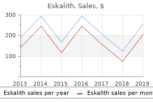
Co-Director, Stony Brook University School of Medicine
Acute inlammatory diseases of the skin tropical depression definition buy eskalith now, commonly lasting a few days to several weeks mood disorder 6 year old boy cheap eskalith 300mg amex, are numerous and pathogenetically inhomogeneous depression definition dsm 5 discount eskalith 300mg on-line. Like other dermatologic diseases anxiety or depression 300mg eskalith fast delivery, they are classiied according to the location of changes. Consequently, there are several classiication schemes for dermatologic diseases, and the examples in this chapter do not favor any particular one. Chronic inlammatory diseases of the skin (chronic inlammatory dermatoses), lasting for several months to years, are also pathogenetically inhomogeneous but have in common signs of chronic inlammation with thickening of the skin, keratinic scale formation, and desquamation (shedding). Among them is systemic lupus erythematosus, which because of its complexity cannot be described in detail here but is discussed in texts on clinical immunology and immunopathology. Vesicular and bullous dermatoses are characterized by epidermal or dermal-epidermal separation (dyshesive diseases) with formation of vesicles or bullae (blisters). They are classiied according to the site where blisters form: epidermolytic blisters comprise supericial intradermal or acantholytic separation and suprabasal epidermal separation, junctional blisters are found below the basal cell layer and dermal lamina densa, and dermolytic blisters form in the upper subepithelial dermis. Although the etiology of these diseases remains obscure, most if not all bullous dermatoses seem to have an autoimmune pathogenesis. There exists a large variety of dermatologic infections, the pathologic changes of which are similar to those in other organs and tissues (taking into account the structural peculiarities of the site of infection): neutrophilic inlammation with or without necrosis and hemorrhage (bacteria, fungi), lymphoplasmacytic iniltration (virus or autoimmune component), and granulomatous (intracellular organisms, fungi, parasites, or autoimmune component). Lesions may be localized (site of entry, lymphatic spread) or systemic (hematogenous spread), occur in supericial or deep parts of the skin, and are usually painful. Infection with certain organisms (Candida, zoster and other herpes viruses, papilloma virus, and others) may suggest that the patient has immune deiciency (opportunistic infections). Hyperplastic changes of the skin consist primarily of hyperplastic scars (keloids) or hyperplastic glands (sebaceous hyperplasia). Benign tumors of the skin are common, and only exceptional cases of conditions such as actinic keratosis (AK) and some forms of nevi progress to malignancy. Benign tumors are usually derived from surface epithelium (seborrheic keratosis, keratoacanthoma, epithelioma of Malherbe), ductular cells of skin appendages (sebaceous adenoma, syringoma, various cysts), or neuroectodermal cells (nevi). Some benign tumors are associated with viral infections (verruca vulgaris, molluscum contagiosum). Benign mesenchymal tumors in the dermis include hemangiomas, lymphangiomas, and ibromas, including skin tags (dermatoibroma, ibrous histiocytoma), neuroibromas, and lipomas. The most frequent malignant tumors of the skin are epidermal neoplasms (squamous cell carcinoma [SCC], basal cell carcinoma [BCC]), melanocytic tumors (malignant melanomas [MMs]), and malignant lymphomas. Less frequent are tumors of the skin appendages (sebaceous cell carcinoma, sweat gland carcinoma), other neuroectodermal tumors (malignant neuroectodermal tumor, Merkel cell tumor), and mesenchymal neoplasms (ibrosarcoma, liposarcoma, hemangiosarcoma, lymphangiosarcoma). Acute eczematous dermatitis is characterized by erythema, edema, and vesicle formation. The etiopathogenesis is similar to urticaria (IgE-dependent immune reaction), but microscopy shows distinct eosinophilia and epidermal spongiosis. Fungal infection (dermatophytosis) must be excluded in the differential diagnosis. Urticaria (hives), an acute pruritic disease of short duration, is caused by a type I immune reaction (allergic, IgE-mediated) that results in local accumulation and degranulation of mast cells with histamine release and edema (pruritic nodule, edematous swelling and plaques, formation of bullae). Urticaria is caused by substances such as drugs, household stuffs, insect bites, and foods that elicit a type I immune reaction. Skin eruption is frequently preceded by malaise, fever, and itching or burning sensations. Typical skin changes consist of target lesions with centrifugal growth, dusky red plaques, and macules and papules on the feet and the extensor surfaces of the arms and legs. Individual lesions heal within 1 to 2 weeks and show variable hyperpigmentation and hypopigmentation. Erythema multiforme (EMF), a common hypersensitivity syndrome with associated vasculitis, may coincide with other diseases. EMF may occur after the administration of certain drugs (sulfonamides, barbiturates, salicylates) or may accompany malignant diseases (carcinoma, lymphoma) and collagen-vascular disorders. Psoriasis is a chronic inlammatory dermatosis of epidermis and dermis with epidermal hyperplasia and hyperkeratosis and parakeratosis. A deregulated epidermal cell proliferation with disturbed microcirculation has been hypothesized. Skin lesions are large (4-5 cm), demarcated, pink plaques with silver-white keratotic scales showing pinpoint hemorrhages (Auspitz sign). Microscopy shows epidermal thickening with elongated rete pegs (acanthosis), loss of the stratum granulosum and parakeratosis, thinning of suprapapillary plates with hyperemic vessels in dermal papillae, and a mixed cellular subepidermal-epidermal inlammatory iniltrate. LP affects skin and mucous membranes and consists of small (2-10 mm) polygonal, white to pink, lat pruritic papules with a crisscrossed surface (Wickham striae). Externally, the papules are located on the lexor surfaces of the wrists, arms, and legs; internally, they appear on the tongue and buccal mucosa as nonerosive or erosive plaques. Microscopy shows liquefaction degeneration of basal cells with subepithelial lymphocytic iniltration. Rete pegs are elongated with hyperparakeratosis, issures, and single cell keratinization (Civatte bodies). Circular vesicles that imitate herpes simplex virus infection is seen in dermatitis herpetiformis. Blister formation via acantholysis is the hallmark histological finding in pemphigus vulgaris. It occurs everywhere on the skin except the palms and soles, with blisters that can be laterally dislocated (Nikolsky sign). Microscopy shows vesicle formation within the stratum spinosum with acantholytic epithelial cells and few lymphocytes, macrophages, or eosinophils. Serum IgG autoantibodies against desmoglein III (intercellular desmosomal component) can be demonstrated.
Perineural invasion is probably American men older than 50 years have a lifetime risk of clinical carcinoma of the prostate of approximately 10% anxiety books purchase eskalith master card. This malignant tumor typically originates in the posterior lobe of the gland anxiety 1-10 scale 300mg eskalith sale, with or without coexistent benign hyperplasia mood disorder related to pms 300 mg eskalith fast delivery. Because androgens stimulate the growth of normal and neoplastic prostatic epithelium depression symptoms worsening cheap eskalith amex, carcinogenesis likely relates to an imbalance in production of androgens and estrogens. However, of utmost importance for cure of prostatic cancer is early detection and treatment before the malignancy has extended beyond the prostatic capsule to involve adjacent pelvic structures or spread to distant sites. Prostatic cancer extends initially by contiguous spread to the bladder and surrounding tissues. It spreads eventually to distant sites by invasion of the bloodstream and lymphatics. Acute hydrocele typically occurs secondary to trauma, tumors, or infection of the testicle and epididymis, particularly gonorrhea and tuberculosis. A spermatocele is a cyst within the scrotum that develops due to obstruction of the sperm transporting system. Rarely, enlargement of the spermatic cord is due to a malignant tumor, typically some type of sarcoma. Hydrocele, an accumulation of serous luid in the 2 peritoneal layers of the tunica vaginalis, results from abnormalities in the descent of the testicles from the retroperitoneal position in the abdominal cavity to the scrotum. The common simple hydrocele is a distended, luid-illed segment of a normally formed tunica vaginalis. Congenital hydrocele, with or without hernia, involves a communication with the abdominal cavity. Torsion (twisting) of the spermatic cord results in compression of the vasculature followed by infarction or complete gangrene of the testicle. Excessive mobility of the testis due to various developmental anomalies is the common predisposing factor. Torsion of the tiny vestigial appendix testis may cause acute pain in the scrotum, which can simulate acute epididymitis or can even mimic acute appendicitis from a referred pain pattern. Varicocele, a collection of dilated, tortuous veins of the pampiniform plexus in the scrotum, is nearly always left sided and asymptomatic. The sudden onset of varicocele after the age of 30 years is secondary to retroperitoneal disease, such as tumors, hydronephrosis, or vascular anomalies. Abrupt onset and rapid progression of gangrene in apparently healthy individuals are initiated by an occult infection (idiopathic or Fournier gangrene). Reduced air circulation and evaporation of sweat in a tight space, irritation of scrotal skin by rubbing against adjacent structures, and ready access to bacteria are common causes of infection in the scrotum. Furuncles develop from hair follicles or sweat glands infected with Staphylococcus aureus. Scrotal erysipelas, a widespread supericial infection of scrotal skin, is usually caused by Streptococcus pyogenes. Eunuchism and eunuchoidism are, respectively, the absence of the testis and severe reduction of androgen production. Primary testicular failure, which typically begins in the prepubertal or very early pubertal period, is the result of various intrinsic developmental defects in the testis with intact pituitary function. This picture is characterized as primary or hypergonadotropic eunuchoidism or hypergonadotropic hypogonadism. Klinefelter syndrome, a genetic disease with a sex chromosomal abnormality, usually XXY, presents at puberty with small testes with hyalinized seminiferous tubules and features of eunuchoidism. The phenotype may be eunuchoid with a speciic pituitary gonadotrophic deiciency or that of a pituitary dwarf with panhypopituitarism. Many patients present with hybrid features of both primary and secondary hypogonadism. Testicular deiciency is secondary to failure of the pituitary to produce gonadotrophic hormones (secondary hypogonadotropic hypogonadism) in approximately 80% of hypogonadal male patients. Although the exact cause of the hypofunction is often unknown, it is important to rule out a pituitary tumor (chromophobe adenoma) or a hypothalamic tumor or other intracranial lesion. The endocrine type (pseudosexual precocity) results from an adenoma, carcinoma, or hyperplasia of the adrenal cortex or an interstitial cell (Leydig cell) tumor of the testis. These patients can combine the macrogenitosomia and premature musculoskeletal development with Cushing syndrome, hypertension, or both. Sexual precocity involves not only premature enlargement of the penis, pubic hair, and testes (macrogenitosomia), but also growth of the skeleton, muscles, body hair, and other structures. Sexual precocity may result from premature development or abnormalities of the hypothalamus and pituitary. Histology of the testis shows a mature pattern with signiicant development of both tubules and interstitial cells. The source of organisms can be infected urine, prostate, or seminal vesicles, with spread via the vas deferens to the epididymis. It may also result from systemic infections or be caused by bacterial toxins from distant localized infections, such as tonsillitis, sinusitis, or cellulitis. Acute pyogenic orchitis (epididymoorchitis) may progress to involve the testis, resulting in a large abscess. Composed of uniform cells with single, central nuclei arranged in solid clusters Embryonal carcinoma Tumor with cystic and hemorrhagic foci.
The clinical presentation of patients with APS I may be impacted by the presence of hypoparathyroidism as well anxiety 5-htp discount eskalith on line. In addition anxiety from alcohol order 300mg eskalith free shipping, observation may reveal opaque depression symptoms help discount 300mg eskalith fast delivery, brittle nails with longitudinal grooves as well as sparse hair depression in adolescence purchase eskalith 300mg online. In addition to the management of endocrine abnormalities, treatment of the cutaneous features of APS employs the use of antifungal agents. Genetic Syndromes Carney Complex the characteristic cutaneous manifestations of Carney complex are spotty skin pigmentation (lentigines) most often found around the lips; on the eyelids, canthi, and the conjunctivae; the ears; and the genital area and present by the time of puberty. Mucocutaneous myxomas, which are derived from effects on fibroblasts, tend to develop within the first two decades of life, are generally asymptomatic sessile or pedunculated pink papules, and are distributed on the eyelids, nipples, buttocks, external ear, tongue, palate, and perineums (Saggini and Brandi 2011). Patients may develop multiple blue nevi (including the epithelioid histologic subtype characteristic of this condition) which have a predilection for acral skin, the buttocks, and the head. The milder enzyme deficiency was termed nonclassical 21-hydroxylase deficiency in 1979 and was later found to be the most common autosomal recessive disorder in humans. Similar to classical congenital adrenal hyperplasia, nonclassical 21-hydroxylase deficiency may cause premature development of pubic hair, advanced bone age, accelerated linear growth velocity, and diminished final height in both males and females. Severe cystic acne has also been attributed to nonclassical congenital adrenal hyperplasia. In males, early beard growth, acne, and growth spurt may prompt the diagnosis of 21-hydroxylase deficiency. Neuroendocrine Tumors Glucagonoma Syndrome Glucagonoma syndrome is a rare disease resulting from a nearly always malignant pancreatic tumor. The distinctive skin lesions of necrolytic migratory erythema are situated mainly on the face in perioral and perinasal distribution, perineum, genitalia, shins, ankles, and feet. Typically, the lesions present as patches of intense erythema that may be angular, annular, or irregular in outline followed by the development of flaccid vesicles and bullae with subsequent denudation of the skin, with crusting and fissuring. Central clearing then occurs, leaving bronze-colored, indurated areas centrally, with blistering, crusting, and scaling at the borders. The cutaneous eruptions occur mainly in the groin, extremities, thighs, buttocks, and perineum. Necrolytic migratory erythema may be complicated by secondary cutaneous infections with yeast such as Candida albicans or bacteria such as Staphylococcus aureus. Skin biopsies obtained from the edge of the lesions reveal superficial necrolysis, with separation of the outer layers of the epidermis, and perivascular infiltration with lymphocytes and histiocytes. In addition, patients may develop glossitis (red, shiny smooth tongue), angular cheilitis, anemia, venous thromboses, and alopecia. Therapy consists in surgical removal of the pancreatic tumor; a near-complete resolution of the skin disease is seen often only 1 week after surgery. Carcinoid Syndrome Most carcinoid tumors originate from the small intestine; they can spread to large parts of the intestinum, the stomach, and the bronchus and may metastasize to the lymph nodes and liver. The skin presents with a pellagra-like appearance lesions with pigmentation and hyperkeratosis on legs and arms. Other cutaneous features include exuberant and transient bright red flushing initially on the face, neck, and upper chest; on repeated episodes, it may involve the entire torso and extremities. Patients may complain of sensations of heat, stiffness, and paresthesias during the attacks. Finally, after prolonged and repeated attacks, a permanent cyanotic flush of the face with telangiectasias resembling rosacea may develop. Merkel cell polyomavirus is thought to be a major contributor to the pathogenesis of the malignancy; however, the exact mechanism of tumorigenesis is unclear and is likely multifactorial. There are several postulated mechanisms, including integration of viral DNA, expression of T-antigen, and evasion of host immune response under the influence of exposure to ultraviolet radiation. Clinically, Merkel cell carcinoma occurs on the head and neck, with predilection for the eyelids and periorbital skin as well as extremities. The tumor may present with various skin manifestations but typically develops as a small, less than 2 cm painless, violaceous papule, plaque, cyst, or infiltrative nodule. Giant variants, mucosal involvement, ulceration, and multiple tumors are less commonly observed. Histopathology shows a tumor of small blue cells with hyperchromatic nuclei and scant cytoplasm, which frequently extends into the subcutaneous fat. Treatment includes surgery with reconstruction associated with radiotherapy and/or chemotherapy for advanced stages including widespread metastases (Daoud et al. Summary Cutaneous lesions can serve as a marker of endocrine disease; it is important for clinicians to be aware of their importance for a prompt and adequate diagnostic approach to the patients. A common feature of these conditions is the secretion of hormones or bioactive substances which produce both widespread multisystem effects and prominent cutaneous signs. In this chapter, the main skin diseases having an endocrinological basis are described. These include cutaneous features of diabetes mellitus, diseases due to alterations of corticosteroid and androgen metabolism, diseases due to dysfunctions of growth hormone secretion, diseases due to dysfunctions of the thyroid gland, paraneoplastic syndromes, and genetic syndromes including dermatological and endocrine symptoms. Acromegaly is associated with decreased skin transepidermal water loss and temperature, and increased skin pH and sebum secretion partially reversible after treatment. New and established topical corticosteroids in dermatology: clinical pharmacology and therapeutic use. Epidermolysis bullosa acquisita in a patient with multiple endocrinopathies syndrome. Multiple facial angiofibromas and collagenomas in patients with multiple endocrine neoplasia type 1.
Chlorhexidine gluconate or tetracycline mouthwash anxiety yoga exercises generic 300 mg eskalith fast delivery, dapsone mood disorder 4 year old purchase eskalith 300mg free shipping, colchicine depression test best best purchase for eskalith, thalidomide and azathioprine have all been used with variable effect depression mental health definition eskalith 300mg fast delivery. Oralpigmentedlesions Non-neoplastic lesions Racial pigmentation is scattered and symmetrically distributed. Amalgam tattoo is the most common form of localized oral pigmentation and consists of blue-black macules involving the gingivae and results from dental amalgam sequestering into the tissues. Neoplastic lesions these include melanotic naevi on the hard palate and buccal mucosa. Malignant melanomas are rare, more common in males and occur mainly on the upper jaw. Neoplasia(squamouscellcarcinoma) Malignant tumours of the mouth account for 1% of all malignant tumours in the UK. Early lesions may be painless, but advanced tumours are easily recognizable as hard indurated ulcers with raised and rolled edges. Premalignant lesions include leucoplakia (single adherent white patch), lichen planus, submucous fibrosis and erythroplakia (a red patch). The tongue the tongue may be affected by inflammatory or malignant processes with similar lesions to those described above. It is also seen in infections due to Candida and in riboflavin and nicotinic acid deficiency. A black hairy tongue is due to a proliferation of chromogenic microorganisms causing brown staining of elongated filiform papillae. The causes are unknown, but heavy smoking and the use of antiseptic mouthwashes have been implicated. There are erythematous areas surrounded by welldefined, slightly raised irregular margins. The gums the gums (gingivae) are the mucous membranes covering the alveolar processes of the mandible and the maxilla. Treatment is with oral metronidazole 200 mg three times daily for 3 days, improved oral hygiene and chlorhexidine gluconate mouthwash. Desquamative gingivitis is a clinical description of smooth, red atrophic gingivae caused by lichen planus or mucous membrane pemphigoid. Salivary duct obstruction due to calculus Obstruction to salivary flow is usually due to a calculus. There is a painful swelling of the submandibular gland after eating and stones can sometimes be felt in the floor of the mouth. Plain X-ray films and sialography will show the calculus; removal of the obstruction by sialoendoscopy gives complete relief. The main cause in man is Streptococcus mutans, which is cariogenic only in the presence of dietary sugar. Dental caries can progress to pulpitis and pulp necrosis, and spreading infection can cause dentoalveolar abscesses. The pleomorphic adenoma is the most common and 15% of these undergo malignant transformation. Malignant tumours classically result in lower motor neurone 7th cranial nerve signs. In the upper portion of the oesophagus, both the outer longitudinal layer and inner circular muscle layers are striated. In the lower two-thirds of the oesophagus, including the thoracic and abdominal parts containing the lower oesophageal sphincter, both layers are composed of smooth muscle. The oesophagus is separated from the pharynx by the upper oesophageal sphincter (UOS), which is normally closed due to tonic activity of the nerves supplying the cricopharyngeus. Oral manifestations of HIV infection HIV-infected patients often have characteristic oral lesions. Swallowing During swallowing, the bolus of food is voluntarily moved from the mouth to the pharynx. This process is mediated by a complex reflex involving a swallowing centre in the dorsal motor nucleus of the vagus in the brainstem. Once activated, the swallowing centre neurones send pre-programmed discharges of inhibition followed by excitation to the motor nuclei of the cranial nerves. This results in initial relaxation, followed by distally progressive activation of neurones to the oesophageal smooth muscle and LOS. Pharyngeal and the principles of management are to preserve what flow remains, stimulate flow and replace saliva (glycerine and lemon mouthwash and artificial saliva). Bacterial sialadenitis is a painful ascending infection with the pharynx and oesophagus Dorsal motor nucleus of the vagus Table6. Odynophagia is pain during the act of swallowing and is suggestive of oesophagitis. Causes include reflux, infection, chemical oesophagitis due to drugs such as bisphosphonates or slow-release potassium or associated with oesophageal stenosis. It is usually a retrosternal burning pain that can spread to the neck, across the chest, and when severe can be difficult to distinguish from the pain of ischaemic heart disease. It is often worst lying down at night when gravity promotes reflux or on bending or stooping. Regurgitation is the effortless reflux of oesophageal contents into the mouth and pharynx. Uncommon in normal subjects, it occurs frequently in patients with gastro-oesophageal reflux disease or organic stenosis.